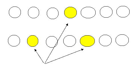
Antibody-dependent cytotoxicity
Mechanism: Either IgG
or IgM is made against antigens. The binding of these
antibodies to the surface of host cells then leads to:
1.
opsonization
of cells whereby phagocytes stick to host cells by way of IgG,
and discharge their lysosomes and ;
2.
activation of the classical
complement pathway causing lysis of the cells
3. ADCC
destruction of the cells whereby
NK cells attach to the Fc portion of the antibodies. The NK cell
then release pore-forming proteins called perforins and proteolytic enzymes
called granzymes. Granzymes pass through the pores and activate the enzymes
that lead to apoptosis of the infected cell by means of destruction of its
structural cytoskeleton proteins and by chromosomal degradation.
Complement Fixation
The complement fixation assay can be used to look for the presence of i)
specific antibody or ii) specific antigen in a patient's serum. The test
utilizes sheep red blood cells (SRBC), anti-SRBC antibody and complement,
along with specific antigen (if looking for antibody in serum) or specific
antibody (if looking for antigen in serum). If antibody (or antigen) is
present in the patient's serum, then the complement is completely utilized
and SRBC lysis is minimal. However, if the antibody (or antigen) is not
present in the patient's serum, then the complement binds anti-SRBC antibody
and lysis of the SRBCs ensues. The following graphics illustrate the
complement fixation assay.
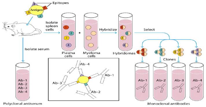
Monoclonal
Ab Production
Balb/c mice
Nuclear
hybridization to give one single nucleus
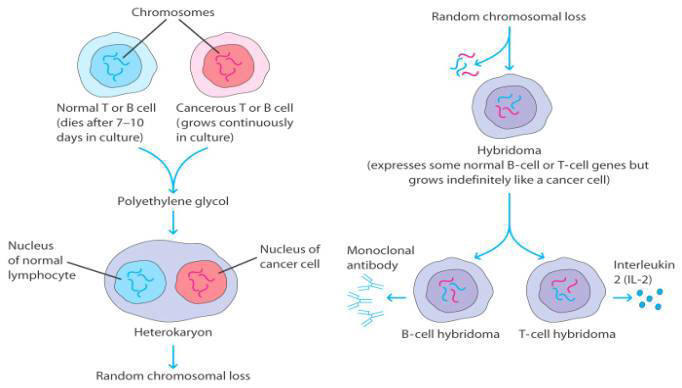
Benefits of using hybridomas:
- Possess
the immortal growth qualities of the myeloma cell
- Secrete
large amounts of monoclonal Ab
- Can
be cultured indefinitely
How to make a M.C. Ab response to blood
Cytochrome
C
- Take
Cyto C
- Inject
into mouse
-
Take
spleen B cells
-
Use
myeloma cell to fuse with B cell (Ab)
-
Bring
together with fusion agent PEG (polyethylene glycol)
What do you want from this?
- You
want to separate the hybridomas
- You
want Ab to Cyto C
To do this:
- You
have to kill off the myeloma cells (use toxic agent)
- B
cells will die on their own (2 weeks)
This will leave you with just the hybridomas
(but a mixture of them)
You then need a selective procedure to pick
out what you need.
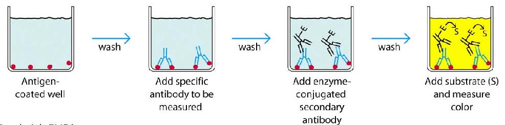
ELISA example
- Screening to see if you have
B
cells that are making Ab to Cyto-C
- Some of the B cells will have
Ab to Cyto-C, some will not.
- Use 96 well plate
- Plate at Conc of 100 cells/well (achieved by
serial dilutions)
- 10 wells
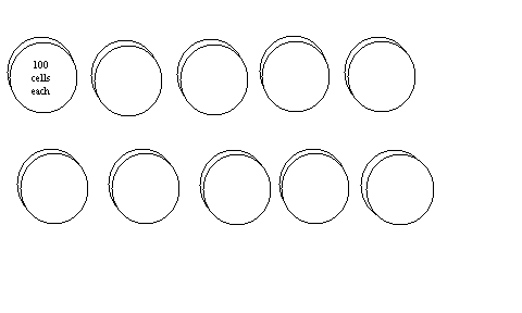
- The anitgen (cyto C) is coated at the
bottom of each well
- If there are Ab to Cyto C in the well
they will attach
- A secondary Ab (which is specific for
the primary {the one for cyto-c}) will bind the primary essentially tagging
it with an enzyme.
- Addition of substrate will cause color
change if the two antibody complex is present
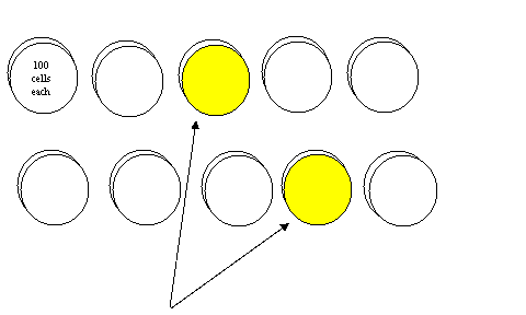
B cells with Ab specific to Cyto-C are present in these two wells
- Now we have narrowed the cells down
from 1000 cells to 200 (100+100)
- Over time you can take these 200 cells
and through serial dilutions split them up so that you eventually have 1 B
cell in every well.
- Again you are using well that are
coated with Cyto-C
- If there is a reaction in that the
well, the B cell clone must be making Ab specific to an epitope on Cyto-C